Now Reading: Kolorektal Karaciğer Metastazında Lenfovasküler İnvazyon Prognostik
-
01
Kolorektal Karaciğer Metastazında Lenfovasküler İnvazyon Prognostik
Kolorektal Karaciğer Metastazında Lenfovasküler İnvazyon Prognostik
Lymphovascular invasion (LVI) has long been recognized as a pathological hallmark associated with aggressive tumor behavior in multiple cancers. Yet, its specific prognostic significance in colorectal cancer liver metastases (CRCLM) remains less well characterized, leaving clinicians and researchers grappling with its therapeutic implications. A recent comprehensive study published in *BMC Cancer* by Li et al. sheds new light on this subject, offering robust evidence that LVI stands as an independent prognostic factor influencing survival outcomes in CRCLM patients undergoing primary surgical resection.
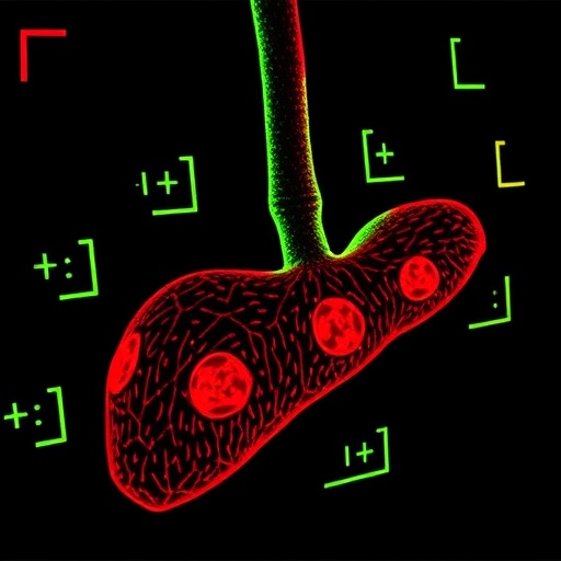
Colorectal cancer frequently metastasizes to the liver, significantly worsening patient prognosis. Surgical resection of liver metastases offers a potential cure, but recurrence rates remain troublingly high. Recognizing additional pathological markers that could predict recurrence and guide adjuvant treatment decisions is a pressing need in the oncology community. The study by Li and colleagues sought to untangle the prognostic relevance of LVI in such metastatic disease, leveraging a large retrospective patient cohort treated at Wuhan Union Hospital, spanning 2013 to 2018.
The researchers applied propensity score matching (PSM), a sophisticated statistical technique designed to minimize confounding by balancing baseline characteristics between groups with and without LVI. This methodological rigor ensured that subsequent survival comparisons and prognostic assessments were not clouded by demographic or clinical disparities often inherent in retrospective analyses. After matching, each group contained 115 patients, establishing a solid basis for unbiased outcome comparisons.
Using multivariate Cox regression models, LVI emerged clearly as an independent predictor adversely affecting both overall survival (OS) and disease-free survival (DFS). Hazard ratios elucidated a roughly 40-45% increased risk of mortality and recurrence in patients exhibiting LVI compared to those without it. This statistically significant finding underscores the aggressive tumor biology associated with lymphovascular invasion, confirming its value as a marker for poor prognosis in CRCLM.
Notably, the study examined the interaction between LVI status and the efficacy of postoperative adjuvant chemotherapy. Intriguingly, while patients without LVI did not show significant survival benefits from chemotherapy, those with LVI experienced markedly improved OS and DFS after receiving treatment. This differential chemotherapy response highlights LVI not only as a prognostic but also a predictive biomarker, identifying patients who may derive substantial benefit from adjuvant systemic therapy post liver resection.
Further substantiating their findings, the authors computed receiver operating characteristic (ROC) curves to evaluate the predictive accuracy of LVI regarding long-term outcomes. The area under the curve (AUC) values for OS and DFS markedly improved in the LVI-positive cohort compared to the entire matched population, particularly at three and five years post-surgery. Such quantitative metrics reinforce LVI’s role in risk stratification, supporting its integration into clinical decision-making algorithms.
This study’s meticulous design and analytical rigor provide a much-needed clarion call for pathology and oncology practitioners to refine pathological reporting standards. Routine evaluation and explicit documentation of LVI in surgical specimens of CRCLM should become standardized practice. Doing so would equip multidisciplinary teams with critical prognostic insights, enabling personalized postoperative management and potentially improving patient survival.
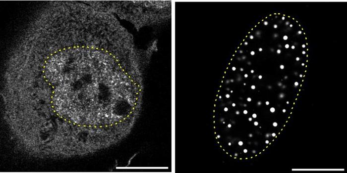
From a mechanistic perspective, lymphovascular invasion indicates cancer cells’ capability to infiltrate local blood vessels and lymphatics, facilitating systemic dissemination. The presence of LVI suggests an early step in metastatic progression, whereby tumor cells breach vascular boundaries, seeding micro-metastases and triggering recurrence even after ostensibly curative resection. Understanding these biological underpinnings can inform ongoing research into targeted therapies addressing metastatic spread at its incipient stages.
The implications of these findings ripple beyond colorectal cancer. LVI has been implicated in breast, lung, and gastric cancers, among others, as a poor prognostic marker. The study by Li et al. contributes to a growing body of literature affirming the centrality of tumor-vascular interactions in cancer progression. It also underscores the importance of adopting integrated approaches combining pathology, surgical oncology, and systemic therapy to optimize outcomes.
Future directions in this domain may include prospective validation of LVI as a biomarker in clinical trials, evaluation of novel agents specifically targeting lymphovascular dissemination, and incorporation of LVI status into existing prognostic nomograms. Moreover, advanced imaging and molecular pathology techniques might enhance the detection sensitivity for LVI, further refining risk assessments.
In conclusion, Li and colleagues have provided compelling evidence that lymphovascular invasion significantly influences the prognosis of patients with colorectal liver metastases following primary tumor resection. Their data reveal that LVI-positive patients not only face worse survival outcomes but also uniquely benefit from adjuvant chemotherapy, emphasizing the marker’s dual prognostic and predictive roles. This landmark study advocates for heightened awareness of LVI in clinical practice, potentially reshaping postoperative management strategies and improving long-term survival for this challenging patient population.
As oncology moves towards precision medicine, the integration of pathological features like lymphovascular invasion with molecular profiling and individualized treatment regimens promises to transform colorectal cancer care. Identifying high-risk patients using robust biomarkers will be key to tailoring therapies, avoiding overtreatment in low-risk groups, and concentrating resources where they can truly impact outcomes. Li et al.’s work marks a significant step forward in that direction, opening avenues for research and clinical application alike.
Such advancements offer tangible hope to thousands worldwide grappling with colorectal cancer liver metastases—a condition historically associated with dismal prognoses. By refining prognostic tools and refining treatment pathways, clinicians can better navigate the complexities of metastatic disease, ultimately translating scientific insight into meaningful patient benefit.
**Subject of Research:**
Colorectal cancer liver metastasis and the prognostic impact of lymphovascular invasion
**Article Title:**
Lymphovascular invasion affects prognosis of colorectal cancer liver metastasis underwent primary resection: a propensity score matching analysis
**Article References:**
Li, W., Liu, B., Xiang, X. et al. Lymphovascular invasion affects prognosis of colorectal cancer liver metastasis underwent primary resection: a propensity score matching analysis. *BMC Cancer* 25, 793 (2025). https://doi.org/10.1186/s12885-025-14083-2
**Image Credits:**
Scienmag.com
**DOI:**
https://doi.org/10.1186/s12885-025-14083-2
**Keywords:**
cancer metastasis prognostic factors, cancer prognosis and treatment, clinicopathologic data, colorectal cancer, colorectal cancer liver metastasis, liver metastasis primary resection, long-term survival colorectal cancer, lymphovascular invasion colorectal cancer prognosis, malignancies lymphovascular invasion impact, postoperative recurrence survival colorectal cancer, propensity score matching analysis, retrospective study colorectal cancer patients, Wuhan Union Hospital research